Pacemaker detection
| Disease area(s): | MRI safety; cardiology; thoracic imaging |
| Data sources: | University Hospitals Plymouth NHS Trust (UHPNT); Royal Cornwall Hospital |
| Project stage: | Dissemination of results |
| Ethical approval: | Granted |
| Principal Investigator: | Dr Mark Thurston |
| Lead Researcher: | Dr Mark Thurston |
| Funder(s): | Royal Cornwall Hospitals NHS Trust |
| Reference: | https://doi.org/10.1007/s10278-022-00663-2 |
Summary
Flagging the presence of cardiac devices such as pacemakers before an MRI scan is essential to allow appropriate safety checks.
This project assessed the accuracy with which a machine learning model can classify the presence or absence of a pacemaker on pre-existing chest radiographs.
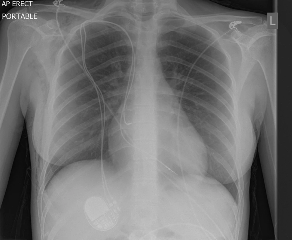
The project analysed a total of 7973 chest radiographs, approximately half the dataset included cardiac devices. The chest X-ray images were reviewed by board-cerfified radiologists to confirm correct labeling. High accuracy (99.67%) was achieved using the machine learning model. This model could be used to screen for patients requiring additional safety input before MRI scan appointments.
The focus of many healthcare applications of computer vision techniques has been for diagnosis and guiding management. This work illustrates an application of computer vision image classification to enhance current processes and improve patient safety.
More information (including the full PDF article) available at Journal of Digital Imaging [open access].